Breast MRI

The breast array coil is an MRI study that offers a complete view of the breasts from any angle. Though mammography is still the most effective way to screen for breast cancer, MRI has gained acceptance in recent years as a secondary tool for diagnosis.
Breast MRI has many benefits: the ability to visualize tumors for staging cancer, to show more detail and detect multiple tumors, to effectively image dense tissue and breast implants, and to capture an expanded view of the chest to determine if cancer has spread to the chest wall or surrounding lymph nodes.
What to Expect:
- Prepare as specified above for a standard MRI.
- You will have two scans: one lying on your back and the second lying on your stomach after receiving a contrast medium. The contrast is given intravenously, and is usually well tolerated. Let your physician know if you have ever had allergic reactions to contrast.
- The actual scan utilizing the breast coil will be the second one, lying face down, as your breasts fit comfortably within the coils on the table. There will be no “squeezing” characteristic of a mammogram. The scans usually take 30 to 45 minutes.
CT (Computed Tomography)

Now more than ever, things move at a faster pace – including the lives of our patients.
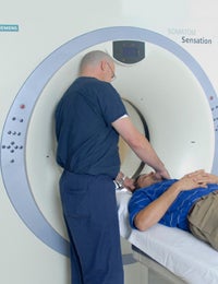
A CT uses X-rays to capture extremely detailed cross-sectional images of your body, called slices. The image slices allow physicians to view internal organs, tissue, bones and blood vessels at various angles. In fact, the technology is so innovative that it can perform CT angiography – non-invasive scan that visualizes blood flow in the arteries and veins.
Our Siemens Somatom Sensation® 64-slice CT is able to capture these images in a breath-hold, which means the exam time is shorter. Depending on the type of scan, a contrast medium may be administered to aid in clarifying the images.
What To Expect:
- You will be asked to remove all jewelry, hairpins, eyeglasses, hearing aids and dentures. For abdominal exams, your doctor should direct how long before the scan you should refrain from eating or drinking.
- Let your physician know if you have had allergic reactions to a contrast medium, iodine or shellfish, or if you have asthma. Also inform the radiologic team if you have diabetes or take medication.
- If contrast medium is used, it will be given either intravenously, or by mouth to coat the gastrointestinal tract. Most people tolerate the contrast medium without any problems and merely feel flushed for a moment.
- Though the images are acquired within a few seconds, the entire exam takes about 15 to 30 minutes – longer if contrast medium is used.
MRI (Magnetic Resonance Imaging)

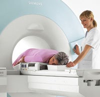
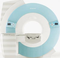
Ochsner Lafayette General Imaging uses a Siemens MAGNETOM EspreeTM short-bore MRI, providing the most patient-friendly, yet powerful open design available today. Using a huge magnet and radio waves, the MRI scan creates a high-resolution image of body tissues.
The open design eliminates the uncomfortable, closed-in “tunnel” of the past, yet facilitates a higher strength magnet than traditional open MRI’s – for superior image quality. With generous amounts of head and elbow room, this unit can be less traumatic for younger and claustrophobic patients since bodily contact is possibly during the scan. From pediatric imaging to exams with the frail elderly, this MRI can handle it all.
What to Expect:
- No special preparation is needed prior to your MRI. Eat normally and take medications as usual, unless your doctor has given you other instructions.
- You will be asked to remove your eyeglasses, watch, jewelry, credit cards, dentures, hearing aids and any other metallic objects you are carrying.
- Generally, you will lie on your back. A lightweight surface coil(s) may be placed over the part of the body to be scanned. The exam itself usually takes 15 to 30 minutes.
PET/CT (Positron Emission Tomography)
The PET/CT is breakthrough technology that combines the imaging capabilities of PET and CT equipment in a single non-invasive procedure. A PET scan detects changes in the chemical functioning of cells in internal organs and living tissue, diagnosing the presence of disease at the molecular level. The CT pinpoints exact location with fine structural detail, superior image quality and a more efficient scan of the anatomy.
For the PET, you will most likely drink or be injected with a drug that contains a dose of radiation to trace the path of the disease. The PET/ CT at Ochsner Lafayette General Imaging is a Siemens BiographTM 16, providing exceptional image quality and complete information regarding the exact location, size, nature and extent of disease, anywhere in the body. PET/CT is especially useful in detecting heart valve viability, Alzheimer’s versus dementia and tumors throughout the body.
What to Expect:
- For six hours before your test, do not eat or drink (except water); do not even chew gum. Your last meal should be high in protein and low in carbohydrates.
- Take medication as prescribed but, if taken with food, try to eat a few soda crackers only four to eight hours prior to your exam. If you have diabetes, call the Ochsner Lafayette General Imaging staff at least 48 hours before your appointment.
- Most people receive the radiopharmaceutical (called FDG) intravenously, a procedure that is usually painless. You will then rest quietly in a comfortable chair for about 60 to 90 minutes to allow the FDG to distribute throughout your body. The scan itself will take between 15 and 40 minutes.
Ultrasound

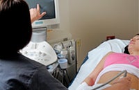
Ultrasound uses high-frequency sound waves to capture images within the body. The Siemens Antares ultrasound unit records and displays the reflected sound wave echoes as images in real time. This imaging system is especially effective at showing the movement of internal tissues and organs, such as the gallbladder, liver, pancreas and kidneys, as well as the flow of blood through the arteries and heart valves.
Of course, ultrasound is best known for fetal imaging — providing parents with that first “picture” of their baby. The technologist at Ochsner Lafayette General Imaging is certified in abdominal and OB/GYN diagnostic medical sonography, with additional experience in what’s called “small parts” tissue like the thyroid and testicles.
The technologist at Ochsner Lafayette General Imaging is certified in abdominal and OB/GYN diagnostic medical sonography, with additional experience in what’s called “small parts” tissue like the thyroid and testicles.
What to Expect:
- Pre-exam preparation will depend on the type of ultrasound, and your doctor will give you specific direction. For some exams, you may need to abstain from eating or drinking for a time prior to the exam; or you may have to drink up to six glasses of water two hours beforehand for a full bladder.
- Wear comfortable, loose-fitting clothing. There are no x-rays or contrast medium involved, and the procedure is virtually painless.
- To perform the exam, the technologist will spread a warm gel on the skin over the area of interest, place a small device called a transducer firmly against the body, and sweep it back and forth to capture the image.


